Surgical removal of the poison glands of rattlesnakes – Tait, 1938
Introduction:
– This publication focuses on the removal of venom glands from a snake via anesthesia with the intention of it returning back to captivity in relatively perfect health. This was to aid in physiological experiments without the fear of being bitten. The procedure in this study used anaesthesia that allowed for a fast recovery to consciousness, but a water-soluble anesthetic could be used like dial or Nembutal which would retain the unconscious state for hours.
Procedure:
– This procedure may be useful to use on the western hognose snakes in my own experiment to retrieve the venom glands.
– The snake is first placed in a glass box filled with chloroform vapour for 20-30min and is laid length wise on a wooden plank and kept in position via strips of plaster. Another method is to drop the snake into a plastic tube where the tail and head is on the open ends and a transverse bar attached to the table keeps the tube from rolling. Next a glass tube pumping oxygen and connected to an ether bottle is inserted into the trachea with a surgical cloth covering the snake except for the head. Anesthesia reactivity is measured using spontaneous body movements, reflex of the tongue when grasped, and reflex of the tail. Preferably spontaneous movements are absent but the other variables are still present, if tongue reflex is absent then it is too much anesthesia. A turgid or full venom gland is easier to locate and isolate compared to an empty one. -> therefore maybe skip feeding on the hognoses prior to euthanizing to retain a larger venom gland. Check the paper for specific instruments needed in procedure.
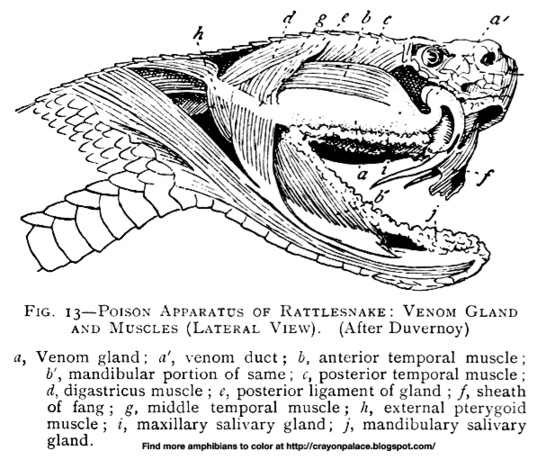
– Pinch the cheek and make a small incision and cut starting slightly above the gape of the mouth to the pit, approximately halfway b/t the eye and mouth. Cut the fibrous tissue underneath the flap which allows for the flaps to be moved upward and downward. The gland is covered in a white fibrous tissue and can be seen when pulling on the downward flap, and the gland ends anteriorly below the eye near the pit where it forms an “S” shape. The posterior end of the gland can be found at the gape of the mouth held by a white band that contrasts the surrounding darker muscle, this will be used to hold onto the gland to begin cutting. The gland is connected to the roof of the mouth via fibers of connective tissue and less dense posteriorly, thus start from the back and move forwards. The posterior end of the gland where the white band is located is also close to important arteries, therefore insert a tool in b/t the band and the other structures so that only the band. The gland is easier to remove from the bottom and lessens the risk of bursting the gland.
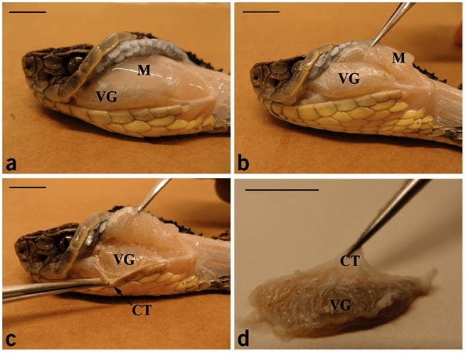
– Next there is a flat compressor muscle along the upper edge of the gland that moves medially and posteriorly and is connected to the gland via ribbon like fibers. Snip these from the posterior end and continue anteriorly. When this is flipped back a horizontal broad ligamentous band can be seen on the medial border of the gland, slip a blade between the ligament and the gland and snip it. At this point the venom gland should resemble a mushroom with only the ventral stalk still connecting it to the head.
– Next tie a catgut string around the ventral stalk and tie it tightly closing the stalk and use scissors to cut above it to free the gland. -> Check the paper for steps on sutchering the wounds.
