Social enrichment reverses the isolation-induced deficits of neuronal plasticity in the hippocampus of male rats – Biggio et al. 2019
Introduction:
Adolescence is a critical life stage for subjects because this is a time when the brain is exhibiting high functions and high amounts of adaption, and if stressed during this time it can cause molecular, structural and functional alterations in the brain. Some of these affect the formation of the hippocampus, specifically stress can cause decreases in Nerve Growth Factor (NGF) and Brain Derived neurotrophic factor (BDNF), reductions in dendritic arborization, spines density and neurogenesis. These changes can cause anxiety, however if subjects are exposed to enrichment, specifically social enrichment then the effects of stress can be reduced. The medial prefrontal cortex, amygdala and hippocampus are 3 brain areas that are responsible for modulating emotions and can be affected by stress. Environmental enrichment is considered one of the best ways to improve brain function through physical and social stimuli. This can include providing toys or ways of increasing physical activity like a running wheel for rodents. Social enrichment has also been shown to increase hippocampal neurogenesis, and aids in involving new neurons into functional circuits that have been harmed by stress.
BDNF and NGF are the most important neurotrophins for brain development and involves neuron survival, specialization, migration, synaptogenesis, spine density, and dendritic arborization, all helping in learning and memory. This study specifically looks at the effects of stress that arises from post-weening isolation by observing BDNF and NGF content, neurogenesis, spine density, and dendritic arborization in the dentate gyrus of rats. Male rats were exposed to 4 weeks of post-weaning social isolation followed by 4 weeks of social housing. They found data that shows the decreases in BDNF, NGF and Arc protein expression as a result of the isolation including reduction in dentate gyrus neurogenesis, spine density and dendritic arborization could be reversed with social enrichment.
Methods:
– Animals = 128 male Sprague-Dawley rats were used for this experiment.
– Social isolation and enrichment = At weaning (PND 21) the rats were separated into 3 groups,1. group housed with 5 animals per cage, 2. socially isolated where it was a individually housed, 3. animals isolated for 4 weeks and then reunited into a group of 5 per cage for 4 weeks.
– Immunoblot analysis = brain was removed and the hippocampus was dissected and weighed. Check the paper for specifics.
– Immunohistochemistry = Neurogenesis and BrdU injection was used to study the proliferation and survival of newborn adult hippocampal neurons. check paper for specifics.
Results:
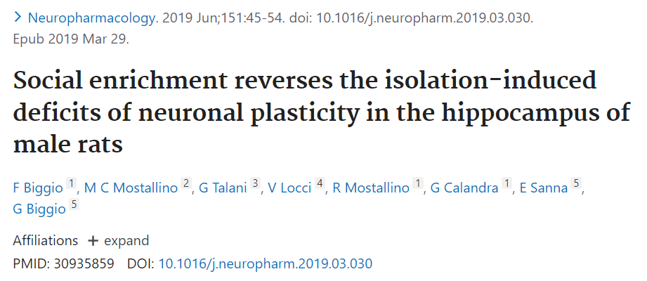
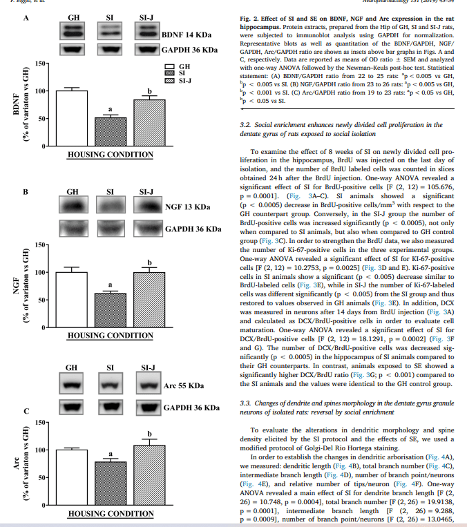
- Social Isolation induces decrease of BDNF, NGF and Arc expression in the rat hippocampus: reversal by social enrichment = BDNF, NGF and Arc protein was observed using immunoblot analysis, there was a significant reduction in all of these parameters when isolated. Then there was a significant increase in all of these parameters when they were returned to social enrichment, these values were similar to the subjects who were group housed from the beginning.
- Social enrichment enhances newly divided cell proliferation in the dentate gyrus of rats exposed to social isolation = After 8 weeks of social isolation, there was a significant decrease of BrdU positive cells when compared with group housed. In the Isolated-social group, they displayed significantly higher levels of positive cells, even more than group housed animals. Additionally Ki-67 labelled cells were observed at significantly lower levels in socially isolated groups compared to group housed and isolated-social group that showed similar values. This shows that the social enrichment reversed the effect of social isolation. A similar case is seen when they observe DCX/BrdU positive cells with social isolation being significantly lower while isolated-social showed similar values to group housed.
- Changes of dendrite and spines morphology in the dentate gyrus granule neurons of isolated rats – reversal by social enrichment = The Golgi-Del Rio Hortega staining was used to observe dendritic morphology and spine density. To understand dendritic arborization they measured 1. dendritic length, 2. total branch number, 3. intermediate branch length, 4. number of branch point/neurons and 5. relative number of tips/neurons. In almost all parameters the socially isolated group was significantly lower than the group housed and isolated-social group except for intermediate branch length in which isolated group was significantly higher. In terms of dendritic spine density, the animals in isolated groups were significantly less dense than group housed and isolated-social. Specifically for spine typologies, isolated rats displayed significantly less mushroom spines compared to group housed and isolated-social group, but thin and stubby spine typologies were at similar levels. Also should be noted that isolated-social group was significantly higher than social isolated but was less than group housed.


Discussion:
Adolescence is a sensitive time for subjects since their brain is in a state of constant development and modification based on surrounding environmental stimuli. Stress at this time can cause lasting mental issues or psychological disorders. Environmental enrichment is a good way to prevent or reverse this, with enrichment being linked with adult hippocampal neurogenesis, integration of new-born cells into functional circuits, and more neuronal plasticity. Social enrichment in particular can have powerful positive effects on brain health and function, with studies showing that rodents living in social enrichment perform better than isolated subjects. Most of the negative effects of social isolation found, like reduction in BDNF, NGF, Arc, neurogenesis of dentate gyrus, spine density, and dendritic arborization were all reversed with social enrichment. The isolated-social group showed values quite similar to the group housed were not significantly different in all parameters. Mushroom spines are involved with long-term potentiation, basically when connections between neurons are made stronger with frequent use, but social isolation causes significant reduction in this typology. With the additional changes in brain formation, social isolation results are similar to subjects with lesions on their hippocampus.
