This is gonna be a bit different than usual posts cause it will be an amalgamation of a couple papers to piece it together for my own experiment with western hognose snakes.
- Evolution and diversification of the toxicofera reptile venom system- Fry et al., 2009
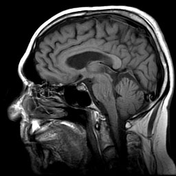
– My experiment will be using western hognose snakes in various environmental conditions to see if it affects cognition and venom. They are hard to extract venom from unless using anesthesia b/c of their weak venom delivery system. One possibility is to use magnetic resonance imaging to see the glands including brain without having to extract it and risk damaging it first.
– The MRI image from this study seems to be really good, it looks like you can see the venom glands relatively clearly as well as other organs in the snake head.

2. Evolution of an arsenal – Fry et al., 2008
This paper takes a more detailed look at comparing the venom glands of different snakes by comparing their toxins, the highlight for my study is a figure they include showing the MRI images of various snakes. It seems like the differences between species is pretty clear visually at this resolution, I wonder if differences b/t individuals of the same species would be discernable as a result of treatment or if the changes resulting from a treatment will need a closer look.
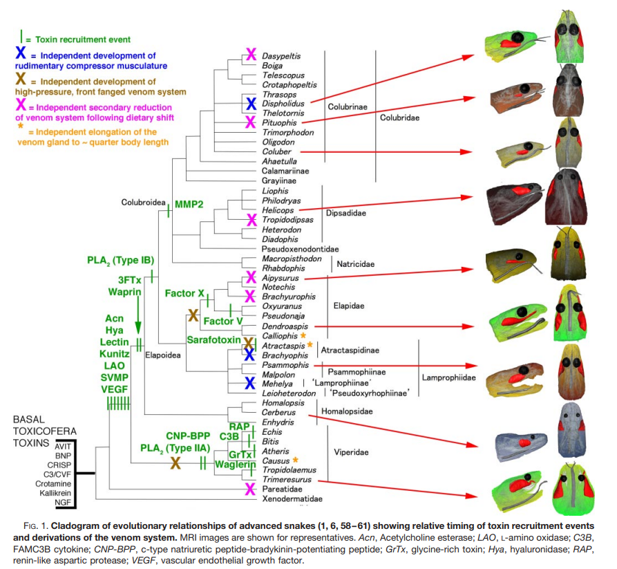
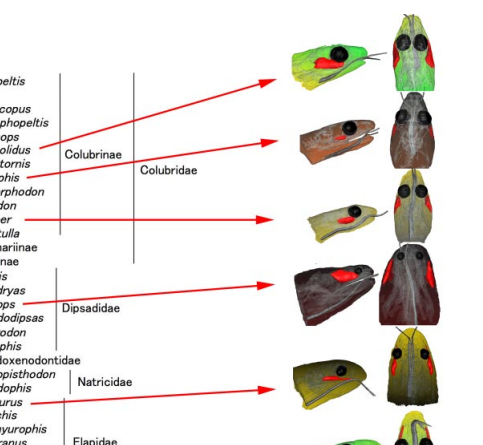
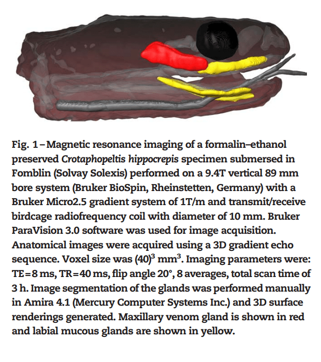
You can see that there are some stark differences b/t species, hopefully this would also be visible b/t individuals. The heads of the subjects were kept in Fomblin and air artifacts were prevented by placing under a vacuum -> check paper for specifics. They also do histological analysis by taking slices of the glands, maybe include this but when a colleague did this it took forever and he had bigger snakes.

